Sunday, June 29, 2008

Compendium Review Unit 3 Major Topic: Nervous Function

Nervous Function

I. Nervous System
II. Senses

I. Nervous System
A. Overview of the Nervous System
1. Functions: receives sensory input, CNS performs integration, CNS generates motor output
2. Nervous tissue: neurons, neuroglia
3. Neuron types and structure

a. sensory neuron, interneuron, motor neuron
b. sensory receptors, effectors
c. cell body, dendrites, axon
4. Myelin sheath
a. formed by Schwann cells in PNS
b. formed by oligodendrocytes in CNS
c. gaps in sheath - nodes of Ranvier
d. gives white, glistening appearance to nerve fibers, good insulator
5. The nerve impulse
a. resting potential - axon not conducting impulse, inside more negative, more Na+ outside, more K+ inside, membrane permeable to K+
b. sodium-potassium pump
c. action potential
i. sodium gates open - Na+ flows in - depolarization: -65mV to +40mV
ii. potassium gates open - K+ flows out - repolarization: 40mV to -65mV
iii. sodium-potassium pump restores resting potential: Na+ out, K+ in
6. Propagation of an action potential
a. each action potential generates another along the length of an axon
b. unmyelinated axon - action potential at 1 locale stimulates adjacent part, 1m/sec
c. myelinated axon - saltatory conduction - 100m/sec
d. multiple sclerosis & leukodystrophies - demyelination - slows propagation
e. all-or-none event
f. intensity of message determined by how many nerve impulses are generated w/in a given time span
g. refractory period - sodium gates cannot open, ensures action potential cannot move backward
7. The synapse
a. axon terminal ends cell body or dendrite of another neuron
b. neurotransmitters transmit impulse across synaptic cleft
i. nerve impulses reach axon terminal
ii. Ca2+ enters terminal - stimulate synaptic vesicles to merge w/sending membrane
iii. neurtransmitter molecules released to synaptic cleft & diffuse to rcving membrane, bind with specific receptor proteins
c. neurotransmitters cause excitation (sodium gates open Na+ in) or inhibition (K+ in)
d. neurotransmitters removed from cleft after initiating response - prevents continuous stimulation/inhibition
e. neurotransmitter molecules
i. ACh, NE, dopamine, serotonin, glutamate, GABA
ii. drugs affecting nervous system: act by interfering w/ or potentiating the action of neurotransmitters
f. synaptic integration - summing of excitatory & inhibitory signals
Figure 13.4 from the text details the structure and fuction of the synapse.B. The Central Nervous System
1. The spinal cord
a. structure
i. gray matter - portions of sensory & motor neurons, interneurons, dorsal root - sensory fibers entering, ventral root - motor fibers exiting
ii. white matter - ascending tracts - info to brain - mostly dorsal, descending tracts - info from brain - mostly ventral
b. functions
i. means of communication btwn brain & peripheral nerves
ii. reflex actions
Figure 13.7 shows an overview of the spinal cord.2. The brain
a. the cerebrum (lateral ventricles)
i. cerebral hemispheres
ii. cerebral cortex - primary motor & sensory areas, association areas, processing centers, central white matter
b. the diencephalon (third ventricle)
i. hypothalamus -integration - maintains homeostasis-regulates hunger, sleep, thirst body temp, water balance. controls pituitary gland
ii. thalamus - rcvs sensory input, integration - visual, auditory, somatosensory, involved w/arousal of cerebrum
iii. pineal gland - secretes melatonin
c. the cerebellum (fourth ventricle)
i. rcvs sensory input - eyes, ears, joints, muslces
ii. rcvs motor output from cerebral cortex - where parts should be
iii. integration - sends motor impulses to skeletal muscles
iv. balance and posture
d. the brain stem - relay station for tracts btwn cerebrum & spinal cord or cerebellum, reflex centers for visual, auditory, tactile responses
i. pons - bundles of axons btwn cerebellum & rest of CNS, works w/medulla oblongata - regulate breathing, has reflex centers - head movement
ii. medulla oblongata - reflex centers - heartbeat, breathing, vasocontriction, vomiting, sneezing, coughing, hiccuping, swallowing
iii. reticular formation - major component of the reticular activating system
Figure 13.10 from the text shows the primary motor and somatosensory areas of the cerebral cortex. Other important images can be found here.C. The Limbic System and Higher Mental Functions
1. The limbic system
a. "evolutionary ancient group of linked structures deep w/in the cerebrum that is a functional group rather than an anatomical one."
b. blends primitive emotions and higher mental functions
c. amygdala - cause experiences to have emtotional overtones
d. hippocampus - learning and memory
2. Higher mental functions
a. memory and learning
i. short-term (prefrontal), long-term (semantic + episodic)
ii. skill memory - involves all motor areas of cerebrum below level of consciousness
iii. long-term memories stored in sensory association areas of cerebral cortex, hippocambus - bridge btwn association areas (storage) & prefrontal area (utilization)
3. Language and speech
i. dependent on semantic memory
ii. seeing & hearing words depends on sensory centers in occipital & temporal lobes
Figure 13.12 from the text illustrates the limbic system of the brain.D. The Peripheral Nervous System
1. Somatic system
a. serve skin, skeletal muscles, tendons
b. nerves - info from external sensory receptors to CNS, motor commands from CNS to skeletal muscles
c. reflexes & the reflex arc - path of nerve impulse when you touch a pin (sensory receptor hand - sensory fibers to dorsal-root ganglia - spinal cord - interneurons - motor neurons - effector )
2. Autonomic system
a. sympathetic and parasympathetic
i. preganglionic fibers arise from middle (thoracolumbar) partion of spinal cord
ii. function automatically and involuntary
iii. innervate all interanl organs
iv. utilize 2 neurons and 1 ganglion for each impulse
b. sympathetic division (NE primary neurotransmitter)
i. preganglionic fibers short, postganglionic fibers long
ii. fight or flight - accelerates heartbeat, dialates bronchi, ihibits digestive tract
c. parasympathetic division (ACh neurotransmitter)
i. few cranial nerves and fibers arising from sacral portion of spinal cord (craniosacral portion of autonomic sys)
ii. preganglionic fibers long, postganglionic fibers short
ii. promotes all internal responses associated with a relaxed state - pupil contraction, promotes digestion, retards heartbeat
Figure 13.14 from the text provides an overview of the PNS. Table 13.1 compares the motor pathways of the somatic and autonomic systems. Click here for images from the text of the somatic system and the autonomic system.E. Drug Abuse
1. Alcohol
2. Nicotine
3. Cocaine
4. Methamphetamine
5. Heroine
6. Marijuana


Definitions from Chapter 13 can be found here.

II. Senses
A. Sensory Receptors and Sensations
1. Types of sensory receptors
a. chemoreceptors
b. photoreceptors
c. mechanoreceptors
d. thermoreceptors
2. How sensation occurs
a. sensory receptors generate nerve impulse
b. if stimulus sufficient, nerve impulse travel along sensory fiber in PNS to CNS
c. nerve impulses reach spinal cord and conveyed to brain
d. if reach cerebral cortex,sensation & perception occur
e. sensory receptors carry out integration b4 initiating nerve impulses (eg sensory adaptation)
Figure 14.1 from the text shows a general overview of sensation and perception. Table 14.1 from the text lists several sensory receptors and related information.B. Proprioceptors and Cutaneous Receptors
1. Proprioceptors
a. mechanoreceptors
b. involved in reflex actions that maintain muscle tone
c. help us know position of limbs in space by detecting degree of muscle relaxation, stretch of tendons, movement of ligaments
2. Cutaneous receptors
a. located in dermal layer of skin, make skin sensitive to touch, pressure, pain, temp
b. touch - Meissner corpuscles, Krause end bulbs, Merkel disks, root hair plexus
b. pressure - Pacinian corpuscles, Ruffini endings
c. temperature - free nerve endings in epidermis
3. Pain receptors nociceptors
a. sensitive to chemicals released by damaged tissues
b. referred pain - eg pain from heart is felt in left shoulder and arm
Figure 14.3 from the text illustrates the various sensory receptors in the skin.C. Senses of Taste and Smell
1. Sense of taste
a. taste buds - on tongue, isolated on hard palate, pharynx, epiglottis
b. how the brain receives taste information - molecules bind to receptor proteins of microvilli on taste cells - nerve impulses generated - travel to brain - interpretation in gustatory cortex
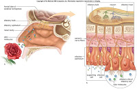
2. Sense of smell
a. olfactory cells - 10 - 20 million high in nasal cavity
b. how the brain receives odor information
i. several hundred types receptor proteins - each olfactory cell has only 1 type
ii. like olfactory cells - nerves lead to same neuron in olfactory bulb
iii. odor molecules bind to specific receptors
iv. odor's signature in olfactory bulb determined by which neurons stimulated
v. neurons communicate info via olfactory tract to olfactory area of cerebral cortex
Figures 14.4 and 14.5 from the text show the taste and smell receptors.D. Sense of Vision
1. Anatomy and physiology of the eye
a. Table 14.2
b. function of the lens
i. cornea with lens and humors - focuses images on retina
ii. viewing near object - ciliary muscle contracts - tension released on suspensory ligaments - allows lens to round up
iii. viewing far object - ciliary muscle relaxed - suspensory ligaments taut - lens is flat
c. visual pathway to the brain
i. photoreceptors - absorption of light ->rod cells - rhodopsin splits into opsin & retinal - release of inhibitory molecules cease - signals go to other neruons in retina. ->cone cells - Blue, green, red pigments
ii. retina - rod & cone cells synapse with bipolar cells - synapse with ganglion cells whose axons become the optic nerve. many (150) rods active 1 ganglion. some cones activates 1 ganglion. integration occuring as signals pass to bipolar & ganglion
iii. blind spot
iv. from the retina to teh visual cortex - impulses from eyes along optic nerve to optic chiasma. fibers from rt 1/2 of each retina converge, continue in rt optic tract. fibers from left 1/2 of each retina converge, continue in left optic tract. fibers synapse with neurons in nuclei w/in the thalamus. nerve impulses to visual cortex
2. Abnormalities of the eye near & farsightedness, astigmatism
Figure 14.6 from the text shows the anatomy of the human eye. Additional images from the text can be found here.E. Sense of Hearing
1. Anatomy and physiology of the ear
a. outer ear - pinna, auditory canal
b. middle ear - tympanic membrane, oval & round window, ossicles (malleus, incus, stapes)
c. auditory tube
d. inner ear - semicircular canals & vestibules (equilibrium) & cochlea (hearing)
e. auditory pathway to the brain - tympanic memb. vibrates - to malleus, incus stapes (pressure multiplied x20). stapes strikes oval window - pressure to fluid in cochlea. movement of pressure waves from vetibular to tympanic canal across basilar membrane causes stereocilia of hair cells to bend. nerve impulses begin in cochlear nerve, travel to auditory cortext in temporal lobe for interpretation
Figures 14.13 and 14.14 illustrate the general anatomy of the ear and, more specifically, of the inner ear.F. Sense of Equilibrium
1. Rotational equilibrium pathway
a. mechanoreceptors in semicircular canals detect rotational equilibrium
b. displacement of cupula in ampulla causes stereocilia of hair cells to bend
c. pattern of impulses to brain changes
d. brain uses info to adjust motor output to right position in space
2. Gravitational equilibrium pathway
a. mechanoreceptors in utricle (back-forth movement) & saccule (up-down movement) detect movement of head in vert or horiz plane
b. movement causes displacement of otoliths, otolithic membrane sags, stereocilia of hair cells bend.
Click here for figure 14.15 from the text which shows the mechanoreceptors for equilibrium.

Definitions from Chapter 14 can be found here.

REFERENCES:
Mader, Syliva S. Human Biology. New York, NY: McGraw-Hill (2008).

Links provided throughout the summary take you to online sources.


IMPORTANT NOTE: Any time "text" or "the text" is referenced in the above summary, I am referring to the textbook Human Biology by Sylvia Mader (cited directly above).

Friday, June 27, 2008

Unit 2 Evaluation

1. What were the three aspects of the assignments I've submitted that I am most proud of?

- The amount of time I spent on the compendiums.
- I like my tables in the lab project.
- Acknowledging that I need to make some changes in my life.


2. What two aspects of my submitted assignments do I believe could have used some improvement?

- I need to spend a little less time on the online labs, and devote more time to the ethical issue.

- I think the conclusion to my lab project could have been better.

3. What do I believe my overall grade should be for this unit?
A

4. How could I perform better in the next unit?
Start the ethical essay and the lab project right away.

Unit 2 Ethical Essay: Food

When I was growing up in my parents house with my 3 sisters, dinner time was an event. It was a group affair. Mom would start dinner. She stayed at home while my dad worked on the farm all day. She made everything from scratch. The four of us would trickle in to help set up the table.

She would yell at us for snacking out of the salad bowl before dinner started. I distincly remember getting my hand slapped for that on more than one occasion. That was and still is one of her biggest pet peeves. Every night we would sit down to the best dinner one of the smallest kitchens (sans T.V.) in existance. We would eat and talk and tease each other.

Occasionally, the teasing would lead into a small food fight. Yes, I am embarrassed to say that I may have missed one of my sisters now and then and regrettably ended up whipping jello on the wall of my parents kitchen. Laughter would erupt; we would get into trouble. We would have to clean the whole kitchen including the walls, by ourselves.

Fast-forward 20 years. My husband and I both work. We both love mountain biking...for the workout, the freedom, the time spent in nature, the smell of the pines, the feel of the air, for the rush. It is a blast. We both feel so lucky to have a hobby that we both love. We can do it together and it is good for us. It gets us outside.

The downside to having jobs and having a hobby that we both like and can spend an endless amount of time doing, is that our time around the dinner table suffers. We tend to make a large portion of something once or twice a week and eat the leftovers for a few days. The good thing is that we love Mexican food. We have probably eaten burritos for dinner 80% of the time over the last year. We are using fairly heathly ingredients (chicken, blackbeans, corn, cilantro, tomatoes, cheese, potatoes), but some of them come from a can instead of being fresh. We also love to have salad every night.

Regardless of what we eat for dinner, we do not spend the time at the dinner table like I did when I was little. It feels like we get home and race through dinner so we can sit down and relax. We need to change our mind set so that we incorporate relaxation into our meal instead of hurrying through it.

I also love the idea of buying locally and eventually growing some of my own produce. I need to figure out how to make that a part of my life. The websites from the link for this essay are a great way to get started.

Unit 2 Lab Project: Exercise Physiology

note: click on any image to view full size

INTRODUCTION
Delivery of oxygen: Possibly the single most important function of the blood that runs through our vessels. We breath air in, the blood in our lungs picks up the oxygen, and after a quick run back through the heart, that oxygenated blood is pumped out to the tissues of the body. The oxygen we breath allows our cells to carry on the never ending cycle of work that they do to keep us alive. Oxygen is part of the final step cellular respiration, the process by which our mitochondria produce ATP, the energy currency of our cells.

The purpose of this lab was to understand the impact that different activities have on basic metabolic rates. Different activities require more or less from our cells. More strenuous activities require more energy; more energy produciton requires more oxygen. The rates we were to measure were pulse, respirations, and systolic and diastolic blood pressures. We were to select 3 different activities to perform. After completing each activity, we were to measure the 4 metabolic rates and compare the mean of each to the mean of our baseline rates.

HYPOTHESIS
HOW DO I THINK MY METABOLIC RATES WILL COMPARE TO BASELINE AFTER....?
My hypotheses are listed in the table below.


MATERIALS AND METHODS
I borrowed a Mabis SmartRead blood pressure monitor from my aunt. This piece of equipment measured systolic blood pressure, diastolic blood pressure, and pulse. The photo below shows the monitor with the cuff wrapped around my bicep. Once the cuff is wrapped tightly around the bicep, there is a blue button that you push. The monitor fulls the cuff with air, slowly releases the air, and provides the readings in the digital display.


To measure my respirations per minute, I used my wristwatch that has a second hand. As the blood pressure monitor was taking my other metabolic measurements, I would keep an eye on my wristwatch and count the number of respirations in 30 seconds. I then took that number and multiplied by 2 to determine the number of respirations per minute.


The first activity I chose was eating. The picture below is of me and my dad getting ready to eat breakfast.


The second activity I chose was a 1 mile time trial on my bike. The picture below is of me riding after I completed the second repetition.


The third activity I chose was a brisk walk around the block. The picture below is of me walking after I completed my first repetition.


DATA
The table below shows the raw data. Included are all four metabolic rates at baseline (5 reps) and after each of the 3 activities (4 reps). Also included is the mean for each.


GRAPH: Mean pulse - baseline and 3 activities


GRAPH: Mean respirations - baseline and 3 activities


GRAPH: Mean systolic blood pressure - baseline and 3 activities


GRAPH: Mean diastolic blood pressure - baseline and 3 activities


ANALYSIS OF DATA
Where my predictions correct?

In the discussion below, when I refer to any of the metabolic rates, I am referring to the mean.

I hypothesized that my pulse would go up as compared to my baseline after each of the 3 activities (eating, biking, walking). I was incorrect when I hypothesized that my pulse would go up after eating. In fact, my baseline pulse and my pulse after eating were the same. My hypotheses about my pulse going up after biking and after walking were both correct.

For respiration rate, I predicted that it would stay the same after eating and go up after biking and after walking. All 3 predictions were correct.

I hypothesized that for systolic blood pressure, each of the 3 activities would cause it to go up. My hypothesis for eating was incorrect. In fact, my baseline and after eating systolic blood pressure were the same. My hypotheses for biking and walking were both correct.

For diastolic blood pressure, I predicted that it would go down after eating and go up after biking and walking. All three of these predictions were correct.


PROBLEMS WITH DATA OR TECHNIQUE
I did encounter a few issues during this experiment.
1. Error message on blood pressure machine - After completing the second repetition of my walk, I tried to measure my blood pressure the monitor gave me an error message. I had to take a second reading which allowed all of my metabolic rates to decrease towards baseline.
2. Time lapse - After my first bike ride, I had to walk into the house to take all 4measurements. Again, this delay allowed my body time to recover. After this I left all necessary equipment in the garage for quick use after biking or walking.
3. Time of day - I took baseline measurements at different times of the day, because I figured my activities would be performed at different times of the day. Ideally, measurements for baseline and all activities would be performed at the same time every day, as baseline metabolic rates vary throughout the day (ie - pulse and blood pressure is lowest in the morning).
4. Measuring respirations - It is very difficult to take this measurement on your own. As soon as you concentrate on counting the number of respirations, you are aware of your breathing rhythm. It is very easy to voluntarily control this rate without meaning to.


CONCLUSION
In general, physical activities are going to increase metabolic rates. These activities consume energy more quickly than sedentary activities do. In response, your body needs to get more oxygen to its cells more quickly. You start breathing harder/faster(respirations) and your heart starts pumping faster (pulse) to move the oxygenated blood to your tissues faster. The faster rate of blood flow causes your blood pressure to rise.

Wednesday, June 25, 2008

Unit 2, Online Lab #2: A Day of Food

My screen shot from the Balance Mind, Body and Soul website.


-How healthy a daily diet do you think this is? Why?
I think this is a fairly healthy diet. It's pretty well-rounded...fruit, vegetables, whole grains, poultry, dairy. It does not include too much processed food.

-What would you change about this day's eating, if anything?
I did not like the sodium intake at all! The saturated fat is higher than I expected as well. This day was somewhat unusual in that I had lunch meat, which I do not normally have. Pasta is not an unusual meal for me, but pasta with sausage is. Those two items really up'd the sodium. Bottom line, I would like to see much lower sodium and saturated fat intake.

-Do you find this kind of nutritional tracking helpful? Why or why not?
I think it's great. In general, I think I have pretty healthy eating habits, but this kind of site lets you know exactly where you stand. What I don't like about this particular site is that (I'm assuming) the nutrition information is based on Sodexo's recipes. I very rarely add salt when I cook. I am assuming again that Sodexo probably prepares their dishes with more salt than I use. This exercise had definitely peaked my interest in nutritional tracking. I would like to use a site that allows me more control of all the ingredients....but at the same time that seems a bit overwhelming! And it might also defeat the purpose. I think the intention here is to give a person a general idea of what kind of diet he or she she has, not to get bogged down with the minutia.

Tuesday, June 24, 2008

Compendium Review Unit 2 Major Topic: Nutrition

Nutrition

Table of Contents
I. Digestive System and Nutrition
II. Converting Food into Energy

I. Digestive System and Nutrition
A. Overview of Digestion
1. Ingestion, digestion, movement, absorption, elimination
2. Wall of the digestive tract
a. mucosa (mucous membrane) - lines tract, protects wall from enzymes, contains glands in mouth, stomach, & small intestine, diverticulitis (pouches in mucosa)
b. submucosa - broad band of connective tissue, contains blood & lymphatic vessels & nerves, inflammatory bowel disease (colitis, inflammatory response)
c. muscularis - 2 layers of smooth muscle (circular & longitudinal), moves GI contents, irritable bowel syndrome (spastic colon, contractions of wall)
d. serosa (serous membrane) - secret serous fluid, part of peritoneum, appendicitis can lead to peritonitis
Figure 8.1 from the text shows an overview of the human GI tractB. First Part of the Digestive Tract
1. The mouth
a. mechanical (teeth and tongue) and chemical (saliva)
2. The pharynx and esophagus
a. swallowing - voluntary and involuntary (once food pushed back into pharynx)
b. peristalsis - rhythmic contraction that pushes food along esophagus
Figure 8.4 from the text shows the path of a bolus as it moves through the first part of the GI tract.C. The Stomach and Small Intestine
1. The stomach
a. continuous with esophagus and duadenum of small intestine
b. stores food, initiates digestion of protein, controls movement of chyme
c. muscularis contains 3 layers of smooth muscle - circular, longitudinal, oblique
d. mucosa has rugae & gastric pits -> gastric glands -> gastric juice (pepsin,HCl, mucus)
2. The small intestine
a. 18ft long
b. digestion completed here
c. duodenum receives enzymes from pancreas and bile from liver and gallbladder
d. nutrients (sugars, amino acids, fatty acids, glycerol) absorbed by sm intestine
e. villi and microvilli increase surface area for absorption
3. Lactose intolerance sufferers lack enzyme, lactase
4. Obesity: diabetes type 2 and cardiovascular disease
a. cells become resistant to insulin and can't utilize glucose
Figure 8.5 from the text illustrates the structure and function of the stomach, table 8.1 which lists the major digestive enzymes, and figure 8.6 which depidcts the anatomy of the small intestine can be found here for later reference. Figure 8.7 which shows digestion and aborption of nutrients is shown below.D. Three Accessory Organs and Regulation of Secretions
1. Three accessory organs
a. pancreas - produces pancreatic juice into duodenum, secrets insulin into blood
b. liver - cleanses blood, stores iron & vitamins, stores glucose, breaks down glycogen, converts glycerol & amino acids to glucose, urea is byproduct, makes plasma proteins
c. gallbladder - stores bile
d. liver disorders - hepatitis, cirrhosis,
2. Regulation of digestive secretions
a. controlled by nervous system and by digestive hormones
Figure 8.8 from the text illustrates the three accessory organs.E. The Large Intestine and Defecation
1. Functions of the large intestine
a. absorbs water
b. intestinal flora produce vitamins which are absorbed by the large intestine
c. forms feces
d. defecation
2. Disorders of the colon and rectum
a. diarrhea, constipation, hemorrhoids, diverticulosis, IBS, IBD, polyps, cancer
Figure 8.10 from the text shows the anatomy of the large intestine.F. Nutrition and Weight Control
1. How obesity is defined
a. body mass index - looks at height and weight
2. Classes of nutrients
a. carbohydrates - simple (eg glucose) complex (body breaks down to glucose)
b. complex carbohydrates better than refined grains
c. proteins - digested to amino acids
d. 8 essential amino acids - daily supply needed
e. lipids - best sources - oils
f. polyunsaturated (contain essential fatty acids)- corn and safflower
g. monounsaturated - olive and canola
3. Minerals
a. trace - body contains less than 5 grams, major - body contains more than 5 grams
b. calcium - construct bones & teeth, nerve conduction, muscle contraction
c. sodium - regulates body's water balance
4. Vitamins
a. 13 vitamins - 4 fat-soluble, 9 water-soluble
b. some are portions of enzymes, others precursors
c. antioxidants - vit. C, E, & A believed to defend the body against free radicals
d. vitamin D - after modification in kidneys & liver, promotes the absorption of calcium by the intestines
5. How to plan nutritious meals
a. eat a variety of foods
b. eat more fruits and vegetables
c. eat less food with saturated or trans fat, sugar, cholesterol, salt, alcohol
d. exercise
e. eat less processed foods
6. Eating disorders
a. anorexia nervosa, bulimia nervosa, binge-eating disorder, muscle dysmorphia
II. Converting Food into Energy
A. Glycolysis1. Glucose converted to pyruvate
2. NET result of single glycolysis run 2 NADH, 2 ATP
3. Lactic acid is end end product under anaerobic conditions
4. 6-Carbon sugar diphospate split into 2 3-Carbon sugar phospate molecules
5. Aerobic conditions, pyruvate is further oxidized to yield more ATP

B. Krebs Cycle1. Occurs in mitochondrion
2. Acetyl-CoA from pyruvate enters Krebs Cycle
3. Single turn of cycle yields 1 ATP, 3 NADH, 1 FADH2
4. Initial reaction involves addition of a 2-Carbon to a 4-Carbon molecule

C. Electron transport chain
1. Electrons are accepted in the following order: cytochrome c, cytochrome c oxidase, oxygen
2. Electrons transferred through the chain originally belonged to NADH and FADH2
3. Movement of protons through ATP synthase: from intermembrane space into matrix
4. Water is produced when oxygent accepts electrons
5. Oxidative phosphorylation is the production of ATP from ADP plus phosphate. The energy used is derived from the movement of proton from the intermembrane space to the matrix

D. How NAD+ Works
1. Cells obtain energy by oxidizing food molecules
2. Coenzyme NAD is reduced NAD+ + H -> NADH
3. Hydrogen atom consists of a proton and an electron
4. Reduction is the addition of an electron, oxidation is the removal of an electron
5. When one molecule is reduced another must be oxidized
6. NADH serves as an electron carrier that can donate its hydrogen


Definitions for chapter 8 can be found here.
REFERENCES:
Mader, Syliva S. Human Biology. New York, NY: McGraw-Hill (2008).

Links provided throughout the summary take you to online sources.


IMPORTANT NOTE: Any time "text" or "the text" is referenced in the above summary, I am referring to the textbook Human Biology by Sylvia Mader (cited directly above).

Wednesday, June 18, 2008

Unit 2, Online Lab #1: Blood Pressure

I was having difficulty emailing and/or copying from the Virtual Lab website, so I decided to create my own graph using the Create a Graph website you recommended. I also typed the journal questions and answers directly into my blog instead of using their journal because of the same issues. I have included screen shots from the Virtual Lab website to show that my work was performed there.

JOURNAL ENTRIES (pre-lab):
Question 1: State a problem about the relationship of age and gender to blood pressure.
Answer 1: Although age and gender impact blood pressure, there are other factors that impact it as well. Genentics and overall health also play a role in determining blood pressure.
Question 2: Use your knowledge about the heart and the circulatory system to make a hypothesis about how the average blood pressure for a group of people would be affected by manipulating the age and gender of the group members.
Answer 2: As age increases, average blood pressure will increase (for both men and women). Men will have higher blood pressure than women.
Question 3: How will you use the investigation screen to test your hypothesis? What steps will you follow? What data will you record?
Answer 3: I will measure the blood pressure of a group of women and a group of men for each age group.

I will start with the youngest age group and measure a group of women. I will stay with the same age group and measure a group of men. I will move on to the next age group and repeat the process until I have gone through each age group. After the measurement is done for each age group/gender combination, I will record the measurements in the table.

After the measurements are complete for each group, I will also review the medical charts and note hereditary history and unhealthy habits that may impact blood pressure.

DATA TABLE AND GRAPH:
The data table is shown in the following two screen shots.


I graphed the data using the Create a Graph website. The first graph shown below is the one from that website. The second graph is a screen shot of the graph generated in the Virtual Lab website. I was not happy with this graph. It was difficult to see the entire graph in one screen shot and the axes were labeled incorrectly.


JOURNAL ENTRIES (post-lab):
Question 4: Analyze the result of your experiment. Explain any patterns you observed.
Answer 4:
  • Over the age of 17 and on average, men have higher blood pressure compared to women of the same age group
  • On average, females in the 18-24 age group have lower blood pressure compared to females in the 11-17 age group
  • On average, females in the 25-34 age group have lower systolic blood pressure compared to females in the 11-17 age group. The average diastolic blood pressure of the same two age groups is the same.
  • On average, the blood pressure of males increases as you move up from one age group to the next.
  • Taking both systolic and diastolic blood pressure into account, the smallest change from one age group to the next is from the 18-24 age group to the 25-34 age group. This is true for men and women.
  • The other contributing factors (weight, family history, salt in diet, exercise, and alcohol consumption) did not seem to have much of an impact on the blood pressure of those in the 25-34 age group. In other words, those in this age group could get away with being a few pounds overweight and/or eating a high salt diet without it negatively impacting their chances of having hypertension.
  • The other contributing fators had a much greater impact for those over 35.
  • Being overweight only did not necessarily mean one will have hypertension.
  • When combined with one of the other factors, being overweight greatly increased the chances of having hypertension.
  • There was not one of the other contributing factors was seen in every person with hypertension.

Question 5: Did the result of your experiment support your hypothesis? Why or why not? Based on your experiment what conclusion can you draw about the relationship of age and gender to group blood pressure averages?
Answer 5: The results of my experiment supported part of my hypothesis to a certain extent. I was correct that men would have higher blood pressure compared to women. I was partially correct when I said that as age increased, so would blood pressure. This was not true when comparing the average blood pressure of those in age group 11-17 to the average blood pressure of those in age group 18-24. Both the average systolic and diastolic blood pressure in the 18-24 year olds was lower than that of the 11-17 year olds. The conclusion that I draw is that in general, as you (male or female) get older, your blood pressure is going to increase. And men are going to have higher blood pressure compared to women of the same age.
Question 6: During the course of your experiment, did you obtain any blood pressure reading that were outside of the normal range for the group being tested? What did you notice on the medical charts for these individuals that might explain their high reading?
Answer 6: Yes, I did obtain blood pressure readings that were outside the normal ranges. I noticed that the high blood pressure could be explained by one of the other contributing factors. For example, most of those with high blood pressure were overweight, from just a few pounds up to almost 50 pounds overweight. I also noticed that most of those with high blood pressure also had one or more of the other contributing factors to blame for it. Some of them also had a diet high in salt, others did not exercise, while others consumed alcohol or had a family history of hypertension. There was only one person who was under weight that had high blood pressure. The only contributing factor that person was guilty of was eating a high salt diet. That person was also male.
Question 7: List risk factors associated with the hypertension. Based on your observation, which risk factor do you think is most closely associated with hypertension?
Answer 7: Risk factors associated with hypertension are as follows:
  • Obesity
  • Family history of hypertension
  • High salt diet
  • Lack of exercise
  • Alcohol consumption

I think being overweight is most closely associated with hypertension.
Question 8: What effect might obesity have on blood pressure? Does obesity alone cause a person to be at risk for high blood pressure? What other factors, in combination with obesity, might increase a person's risk for high blood pressure?
Answer 8: Obesity has many negative health implications. But if you think about just the extra weight an obese person has to carry around....Their heart needs to pump enough blood to supply oxygen and nutrients to that extra weight. The additional blood flow required may increase blood pressure. All of the other factors (listed above) in combination with obesity might contribute to high blood pressure. A few other factors not listed are tobacco use and stress.

Tuesday, June 17, 2008

Compendium Review Unit 2 Major Topic: Oxygen/Microbes/Immunity

Oxygen...Microbes...Immunity

Table of Contents
I. Cardiovascular System: Heart and Blood Vessels
II. Cardiovascular System: Blood
III. Lymphatic System and Immunity
IV. AIDS Supplement
V. Sickle Cell Anemia

I. Cardiovascular System: Heart & Blood Vessels
A. Overview of the Cardiovascular System (heart & blood vessels)
1. Circulation performs exchanges
a. circulation of blood - to service cells
b. blood removes waste product from tissue fluid
c. blood delivers oxygen and nutrients to tissue fluid
d. lungs - exchange carbon dioxide for oxygen
e. kidneys - remove waste
f. intestines - where nutrients enter the blood
g. liver - removes poisons, takes up amino acids and returns proteins
2. Functions of the cardiovascular system
a. heart contracts -> blood pressure -> moves blood
b. blood vessels transport - heart, arteries, capillaries, veins, heart
c. capillaries exchange
d. blood flow regulated by heart and blood vessels
3. Lymphatic system
a. collects excess tissue fluid
Figure 5.1 from the text shows a general view of how the cardiovascular system works with the other systems of the body for the exchange of O2, CO2, waste, and nutrients.

B. The Types of Blood Vessels(artery, arteriole, capillary, venule, vein)
1. Arteries: from the heart (endothelium, smooth muscle & elastic tissue, connective tissue)
2. Capillaries: exchange (endothelium, basement membrane)
a. bypassed by arteriovenous shunt
3. Veins: to the heart (less smooth muscle, less connective tissue, thinner)
a. valves - in veins carrying blood against force of gravity
Figure 5.2 from the text shows an overview of the tissues and vessels that make up and surround a capillary bed.C. The Heart is a Double Pump
1. Passage of blood through the heart
a. O2 POOR blood -> sup vena cava and inf vena cava -> right atrium
b. right atrium -> tricuspid valve -> right ventricle
c. right ventricle -> pulmonary semilunar valve -> pulmonary trunk -> 2 pulmonary arteries -> lungs
d. pulmonary veins with O2 RICH blood -> left atrium
e. left atrium -> bicuspid valve -> left ventricle
f. left ventricle -> aortic semilunar valve -> aorta -> body proper
g. atria - thin walled, ventricles - thicker walled (pump blood farther)
2. Heartbeat is controlled
a. internal control of the heartbeat - SA node-atrium->AV node->AV bundle->Purkinje fibers-ventricles
b. external control of heartbeat - medulla oblongata, epinephrine and norepinephrine
3. Electrocardiogram is a record of the heartbeat (PQRST waves)
Figure 5.3 from the text illustrates the external anatomy of the heart. Figure 5.4 from the text illustrates a cross sectional view of the anatomy of the heart.
D. Features of the Cardiovascular System
1. Pulse rate equals heart rate (arterial walls pulse when left ventricle contracts
2. Blood flow is regulated
a. blood pressure moves blood in arteries
b. blood flow - slow in capillaries
c. blood flow in veins returns blood to heart (blood pressure low, velocity high=skeletal muscle pump, respiratory pump, valves)
d. blood pressure dependent on: volume of space involved, # of molecules in space, kinetic engergy of molecules
E. Two Cardiovascular Pathways
1. Pulmonary circuit: exchange of gases
a. CO2 given off and O2 taken into blood at pulmonary capillaries
2. Systemic circuit: exchanges with tissue fluid
a. tracing the path of blood (aorta, proper branch of aorta, region, returning vein, vena cava)
b. coronary circulation - supplies the heart
c. hepatic portal system - connects digestive tract to liver and liver to inf vena cava
Figure 5.10 from the text shows the flow of blood through the pulmonary and systemic circuits. Figure 5.11 from the text shows the systemic circuit and its major arteries and veins.F. Exchange at the Capillaries
1. Blood pressure and osmotic pressure control movement of fluid through capillary wall
2. Arterial end of cap. bed - blood pressure higher, water out
3. Capillary bed - pressures equal, solutes diffues
(CO2, wastes in & O2, nutrients out)
4. Venule end of cap. bed - osmotic pressure higher, water in
5. Excess fluid collected by lymphatic capillaries

Figure 5.12 shows the exchanges that take place at the capillariesG. Cardiovascular Disorders
1. Disorders of the blood vessels
a. hypertension - high blood pressure (atherosclerosis, plaque, thrombus, embolus)
b. stroke, heart attack, aneurysm
c. dissolving blood clots (t-PA, aspirin)
d. treating clogged arteries (bypass surgery, stent, gene therapy)
2. Disorders of the heart
a. heart transplants (left ventricle assist device, total artificial heart)

Definitions from Chapter 5 can be found here.

II. Cardiovascular System: Blood

A. Blood: An Overview
1. Functions of the blood
a. transport - O2, nutrients, CO2, waste, hormones
b. defense - phagocytosis, antibodies, blood clotting
c. regulation - picks up & transports heat, regulates pH, maintains water-salt balance
2. Composition of blood
a. formed elements - red and white blood cells and platelets, produced in red bone marrow
b. plasma
i. water
ii. salts - buffer
iii. organic molecules - glucose, amino acids, urea, plasma proteins
Below is an image take from this website that shows all of the formed elements of blood.B. Red Blood Cells & Transport of Oxygen
1. How red blood cells carry oxygen
a. hemoglobin - globin=proteing containing 4 highly folded polypeptide chains, heme=iron-containing group in center of polypeptide chain
b. iron accepts O2 in lungs, lets go in tissues
c. 1 RBC (has no nucleus) = 280 million hemoglobin molecules, 1 hemoglobin=4 O2
d. RBCs - biconcave shape=greater surface area for diffusion, internal space=O2 transport
2. How red blood cells help transport carbon dioxide
a. 7% dissolved in plasma
b. 25% combines with terminal amino groups of globin molecules of hemoglobin
c. 68% transported as bicarbonate ion in plasma
e. RBCs - lack most organelles, produce ATP anaerobically, no consumption of O2 they carry
3. Red blood cells are produced in bone marrow
a. RBC stem cell divides -> new cells differentiate into mature RBCs
b. constant regeneration - RBCs only live 120 days
c. RBCs destroyed in liver and spleen by macrophages
d. globin -> amino acids, iron -> marrow, remaining heme portion degraded
e. erythropoietin - produced in kidneys, liver, other tissue - stimulates RBC production in stem cells of bone marrow
f. blood doping
4. Disorders involving red blood cells
a. anemia - too few RBCs or low hemoglobin, due to low iron, vit B12, folic acid
b. hemolysis - rupturing of RBCs, sickle-cell disease
Figure 6.3 from the text shows the single file lines of RBCs moving through capillaries, a close up of RBCs, and the four highly folded polypeptide chains of a hemoglobin molecule.C. White Blood Cells & Defense Against Disease
1. Types of white blood cells
a. neutrophil (granular leukocyte) - 1st responders to bacterial infection
b. eosinophil (granular leukocyte) - increase in # during parasitic worm infection or allergic reaction
c. basophil (granular leukocyte) - release histamine during allergic reaction
d. mast cell (granular leukocyte - release histamine during allergic reaction
e. lymphocyte (agranular leukocyte) - T cells and B cells
f. monocyte (agranular leukocyte) - differentiate into macrophages & dendritic cells
2. Disorders involving white blood cells
a. severe combined immunodeficiency disease
b. leukemia
c. infectious mononucleosis
The image below taken from this website is of a white blood cell that is trapping bacterial cells.D. Platelets & Blood Clotting
1. Blood clotting
a. 12 clotting factors plus Ca2+
b. prothrombin activator+prothrombin+Ca2+->thrombin+fibrinogen+Ca2+->fibrin threads
2. Disorders related to blood clotting
a. thrombocytopenia - insufficient number of platelets
b. thromboembolism - dilodged thrombus obstructs blood vessel
c. hemophilia - inherited clotting disorder, deficiency of clotting factor
Figure 6.8 from the text shows the process of clotting, generalized.E. Blood Typing & Transfusions
1. ABO groups
a. type A blood - type A antigen (on surface of RBC) & anti-B antibodies (in plasma)
b. type B blood - type B antigen & anti-A antibodies
c. type AB blood - type A & type B antigens, no antibodies
e. type O blood - no antigens, anti-A & anti-B antibodies
2. Rh blood groups
a. indicates whether person has Rh factor on RBC
b. Rh- individuals do not have antibody to Rh factor, produce them if exposed
Figure 6.9 from the text illustrates the 4 ABO blood groups and associated antigens and antibodies.F. Homeostasis
Figure 6.13 is a great overview of the contribution that each of the body systems makes to the maintenance of homeostasis.

Definitions from Chapter 6 can be found here.

III. Lymphatic System & Immunity
A. Microbes, Pathogens, & You
1. Bacteria
a. single celled prokaryote, no nucleus
b. shapes=bacillus (rod), spirillum (curved), coccus (spherical)
c. anatomy=fimbriae, flagellum, capsule, cell wall, plasma membrane, piluls, plasmid, ribosome, nucleoid
d. examples of infections - strep throat, TB, botulism, food poisoning, gangrene 2. Viruses
a. bridge gap between living and nonliving
b. acellular
c. obligate parasites
d. outer capsid made of protein, inner core made of nucleic acid
e. adheres to a receptor on cell surface, injects nucleic acid
f. uses cells host's enzymes and ribosomes for replication
g. viral genetic material is DNA or RNA
h. examples of infections - colds, flu, measles, chicken pox, polio, rabies, AIDS
i. emerging viruses - transported to new location, change in vector, change in face
3. Prions
a. proteinaceous infectious particles
b. cause a group of degenerative diseases of the nervous system (CJD, mad cow, scrapie)
Figure 7.1 from the text shows the parts of a bacterium. Figure 7.4 from the text shows the parts of a virus.
B. The Lymphatic System
1. Lymphatic vessels
a. capillaries, vessels, ducts -> cardiovascular veins in shoulders
b. capillaries take up excess tissue fluid
c. thoracic duct and right lymphatic duct
d. vessels - have valves and movement of lymph dependent on muscle contraction
2. Lymphatic organs
a. red bone marrow (makes RBCs, WBCs), thymus gland(makes hormones,maturation of T cells) - both primary
b. lymph nodes (filter lymph), spleen (filters blood), lympatic nodules, Peyer's patches- secondary
C. Nonspecific Defenses
1. Barriers to entry 1st line of defense
a. skin and mucous membranes
b. chemical barriers - skin oil, persperation, tears, saliva, acidic - stomach, vagina
c. resident bacteria - normal flora
2. Inflammatory response 2nd line of defense
a. redness, heat, swelling, pain
b. WBCs rush in - neutrophils, cytokines, monocytes (macrophages), lymphocytes
d. protective proteins - complement proteins - interferons, membrane attack complex
Figure 7.9 from the text depicts the inflammatory response. Figure 7.10 from the text illustrates how protective proteins (complement system) work against a bacterium.D. Specific Defenses
1. How specific defenses work
a. respond to antigens
b. primarily responsible - lymphocytes (T cell & B cell)
c. antibody-mediated immunity - B lymphocytes contain antigen specific receptors
d. antigen fits to BCR, B cell undergoes clonal expansion
e. cloned B cells become plasma cells (produce & secrete antibody) membory cells (can fight same antigen later on)
f. structure of antibody - Y-shaped, 5 classes determined by Y structure, variable region form antigen-bodning site
g. cell-mediated immunity - T lymphocytes, TCR requires antigen-presenting cell (APC) which already phagocytized a pathogen & presents antigen to TCR on self protein
h. T cell compares antigen to self protein and activation occurs, clonal expansion
i. cytotoxic T cells (cause apoptosis in virus-infected or tumor cell) & helper T cells (regulate immunity by secreting cytokines) produced
Tables and images from the text have been uploaded for later reference here. Figure 7.12 from the text shows the structure of an antibody. E. Acquired Immunity
1. Active immunity
a. vaccines - non-virulent pathogen or it's product
b. vaccine exposure->initially no antibody->antibody increases->levels off->declines
c. booster - second exposure to increase - titer increase to greater level
2. Passive immunity
a. prepared antibodies or immune cells given to combat a disease
b. example - newborn infants (antibodies from mother thru placenta, breast milk)
c. monoclonal antibodies - detect pregnancy, ID infections, deliver toxic drugs to tumors
d. cytokines & immunity - interferons & interleukins being investigated to use as adjuncts for vaccines and for cancer treatment
Figure 7.17 from the text illustrates the production of monoclonal antibodies.F. Hypersensitivity Reactions
1. Allergies
a. immediate allergic response (caused by IgE antibodies) eg - anaphylactic shock
b. delayed allergic response (initiated by memory T cells) - eg - TB skin test, contact dermititis (poison ivy)
2. Tissue rejection
a. prevention - immunosuppressive durgs, xenotransplantation, lab organs
3. Disorders of the immune system
a. autoimmune diseases - myasthenia gravis, multiple sclerosis, systemic lupus erythematosus, rheumatoid arthritis

Definitions from Chapter 7 can be found here.

IV. AIDS Supplement
A. Origin of & Prevalence of HIV
1. AIDS caused by HIV which infects & destroys cells (helper T & macrophages) of immune systems
2. Originated in Africa
3. Prevalence of HIV

a. of the 38.6 million people infected with HIV, 24.5 million in Sub-Saharan Africa, 8.3 million in Asia, 2 million in North America, Wester and Central Europe,1.6 million in Latin America
B. Phases of an HIV Infection
1. Category A: Acute Phase
a. no apparent symptoms
b. highly infectious
c. CD4 T cell count - has never fallen below 500 cells per mm3 of blood
d. at first, antibodies low count so not detectable
e. after time, body starts producing CD4 T cells like crazy to overcome the destruction of them by HIV
2. Category B: Chronic Phase
a. CD4 count - 499 to 200 cells/mm3
b. symptoms related to poor immune system - yeast infections, cervical displasia, prolonged diarrhea, thick sores on tongue, shingles, fevers, fatigue, cough
c. HIV particles - # is increasing
3. Category C: AIDS
a. diagnosed with AIDS
b. CD4 count below 200
c. 1 or more - 25 AIDS defining illnesses (opportunistic infections)
d. death results from: Pneumocystis jiroveci pneumonia, Mycobacterium tuberculosis, toxoplasmic encephalitis, Kaposi's sarcoma, invasive cervical cancer
C. HIV Structure and Life Cycle
1. Structure
a. 2 single strands of RNA (retrovirus)
b. various proteins
c. envelope - contains embedded spikes - Gp120
d. protection: 3 protein coats (nucleocapsid, capsid, matrix)
e. matrix contains 3 enzymes: reverse transcriptase (catalyst for reverse transcription), integrase (catalyst for integration of viral DNA into DNA of host), protease (catalyst for breakdown of newly synthesized viral polypept. into viral proteins)
Taken from this website, the drawing below shows the structure of the HIV virus..2. HIV life cycle
a. attachment - HIV to plasma membrane of target cell
b. fusion - of HIV to plasm membrane
c. entry - uncoating of capsid and protein coats, RNA, viral proteins released
d. reverse transcription - reverse transcriptase - single stranded RNA -> DNA
e. integration - viral DNA and integrase into nucleus of host cell, host cell DNA spliced, and viral DNA integrated (HIV now called provirus)
f. biosynthesis and cleavage - production of more viral RNA, protease cleavs long viral polypeptide chains
g. assembly - of viral enzymes, capsid proteins, and RNA into viral particles
h. budding - virus gets envelope
3. Transmission and Prevention of HIV
a. transmitted through bodily fluids during sexual contact, needle-sharing, transfusions, birth or breast feeding
b. prevention - abstinence, sex with 1 uninfected partner, use of condom
4. HIV testing and treatment for HIV
a. tests for HIV antibody
b. treatment - available but not without concerns (drug-resistant viruses)
c. drug therapy -HAART - different drugs interfere with life cycle of HIV
d. vacccines - being studied
The image below, taken from this website, shows the life cycle of the HIV virus.V. Sickle Cell Anemia
A. Discovery and Biological Basis
1. Discovered by cardiologist James B. Herrick and his intern Ernest E. Irons
2. Patient's physical symptoms: pain in back, muslces of back, arms, dark urine, fever, weakness, dizziness, shortness of breath
3. Patient's blood test: nucleated and sickle shaped RBCs
4. Biology of RBCs

a. RBCs made up mostly of hemoglobin (Hb)
b. Hb made up of 4 protein subunit with iron atom at center of each
c. each iron atom has affinity for O2
d. Hbs most important characteristic: reversibly bind and release O2 equally
e. Hb binds O2 when oxygen pressure is high and releases it when its pressure is low
f. anemia - signifant decrease in amount of functional Hb
g. sickle cell anemia causes depletion
i. O2 carrying capactiy reduced b/c of molecular changes in sickeled cell
ii. peculiar shape and rigidity, stick together and clog small arteries
h. effects
i. short term: poor O2 delivery causes shortness of breath
ii. long term: oxygen deprivation leads to poor tissue development
iii. hemolysis & clogging of arteries and capillaries in lungs, kidneys, & liver causes system malfuction and death usually by 30
5. Genetic basis of sickle cell anemia
a. 2 forms: sickle cell anemia (more severe) and sickle cell disease (rarely show complications) discovered
b. sickle cell disease - threshold effect, where a quantitative change produces a qualitative change
6. Localization of the genetic defect
a. through experimentation, defect determined to be in Hb molecule
B. Molecular Biology of Sickle Cell Anemia
1. Electrophoresis helped determine details of sickle cell anemia
a. inherited in simple Mendelian fashion
b. incomplete dominance (those with sickle cell trait have half normal RBCs and half sickle cell RBCs)
c. 1st genetic disease localized to a defect in the structure of a specific protein molecule
2. Sickle cell and normal hemoglobin
a. two-dimensional paper chromatography used to determine the peptide fragment of Hb that was different between normal and sickle cell RBCs (Linus Pauling)
b. Vernon Ingram determined the two Hb molecules differ by 1 amino acid
3. Discovering the difference between normal and sickle-cell Hb
a. Hb made up of 2 alpha and 2 beta chain (4 total) polypeptide chains
b. sickle cell Hb has 2 normal alpha chains; mutation in #6 position of both beta chains
c. protrusion formed by substitued aa at #6 location in beta chains locks into complementary site on other beta chains, linking sickle cell Hb molecules
C. Biogeography and Ecology of Sickle Cell Anemia
1. Unique geographic distribution pattern
a. high frequency of sickle cell anemia in families of African descent
b. higher frequency of sickle cell trait in Africa compared to US
c. sickle cell disease in Africa not common - high infant mortality rate for homozygous recessive infants
2. The malarial connection
a. frequency distribution of malaria mapped out closely to that of sickle cell
b. hypothesized in 1954 by Anthony Allison that heterozygous individuals for sickle cell have an advantage in combating malaria over those with normal Hb
c. balanced poymorphism explains how mutant gene (though by itself does not have an advantage) is selected for in the presence of malaria
3. How does sickle cell help combat malaria?
a. asexual reproductive stage of the protozoan consume high amounts of oxygen, as sickle cell become depleted of oxygen, they sickle and are removed from the body by the spleen....along with the merozoites
4. Treatment and Political Aspects
a. continuing search for treatment
b. drug therapies - some success, most limit detrimental effects instead of curing
c. gene Therapy - goal - replace bad gene Hbs with good allele Hba
d. political aspects - less money raised for sickle cell disease compared to predominantly caucasian afflicting diseases (cistic fibrosis and muscular dystrophy)

REFERENCES:
Mader, Syliva S. Human Biology. New York, NY: McGraw-Hill (2008).

The section on sickle cell anemia (roman numeral five) is a summary of the information provided in this website.

Links provided throughout the summary take you to online sources.


IMPORTANT NOTE: Any time "text" or "the text" is referenced in the above summary, I am referring to the textbook Human Biology by Sylvia Mader (cited directly above).