I. Nervous System
II. Senses
I. Nervous System
A. Overview of the Nervous System
1. Functions: receives sensory input, CNS performs integration, CNS generates motor output
2. Nervous tissue: neurons, neuroglia
3. Neuron types and structure
a. sensory neuron, interneuron, motor neuron
b. sensory receptors, effectors
c. cell body, dendrites, axon
4. Myelin sheath
a. formed by Schwann cells in PNS
b. formed by oligodendrocytes in CNS
c. gaps in sheath - nodes of Ranvier
d. gives white, glistening appearance to nerve fibers, good insulator
5. The nerve impulse
a. resting potential - axon not conducting impulse, inside more negative, more Na+ outside, more K+ inside, membrane permeable to K+
b. sodium-potassium pump
c. action potential
i. sodium gates open - Na+ flows in - depolarization: -65mV to +40mV
ii. potassium gates open - K+ flows out - repolarization: 40mV to -65mV
iii. sodium-potassium pump restores resting potential: Na+ out, K+ in
6. Propagation of an action potential
a. each action potential generates another along the length of an axon
b. unmyelinated axon - action potential at 1 locale stimulates adjacent part, 1m/sec
c. myelinated axon - saltatory conduction - 100m/sec
d. multiple sclerosis & leukodystrophies - demyelination - slows propagation
e. all-or-none event
f. intensity of message determined by how many nerve impulses are generated w/in a given time span
g. refractory period - sodium gates cannot open, ensures action potential cannot move backward
7. The synapse
a. axon terminal ends cell body or dendrite of another neuron
b. neurotransmitters transmit impulse across synaptic cleft
i. nerve impulses reach axon terminal
ii. Ca2+ enters terminal - stimulate synaptic vesicles to merge w/sending membrane
iii. neurtransmitter molecules released to synaptic cleft & diffuse to rcving membrane, bind with specific receptor proteins
c. neurotransmitters cause excitation (sodium gates open Na+ in) or inhibition (K+ in)
d. neurotransmitters removed from cleft after initiating response - prevents continuous stimulation/inhibition
e. neurotransmitter molecules
i. ACh, NE, dopamine, serotonin, glutamate, GABA
ii. drugs affecting nervous system: act by interfering w/ or potentiating the action of neurotransmitters
f. synaptic integration - summing of excitatory & inhibitory signals
Figure 13.4 from the text details the structure and fuction of the synapse.
 B. The Central Nervous System
1. The spinal cord
a. structure
i. gray matter - portions of sensory & motor neurons, interneurons, dorsal root - sensory fibers entering, ventral root - motor fibers exiting
ii. white matter - ascending tracts - info to brain - mostly dorsal, descending tracts - info from brain - mostly ventral
b. functions
i. means of communication btwn brain & peripheral nerves
ii. reflex actions
Figure 13.7 shows an overview of the spinal cord.
 2. The brain
a. the cerebrum (lateral ventricles)
i. cerebral hemispheres
ii. cerebral cortex - primary motor & sensory areas, association areas, processing centers, central white matter
b. the diencephalon (third ventricle)
i. hypothalamus -integration - maintains homeostasis-regulates hunger, sleep, thirst body temp, water balance. controls pituitary gland
ii. thalamus - rcvs sensory input, integration - visual, auditory, somatosensory, involved w/arousal of cerebrum
iii. pineal gland - secretes melatonin
c. the cerebellum (fourth ventricle)
i. rcvs sensory input - eyes, ears, joints, muslces
ii. rcvs motor output from cerebral cortex - where parts should be
iii. integration - sends motor impulses to skeletal muscles
iv. balance and posture
d. the brain stem - relay station for tracts btwn cerebrum & spinal cord or cerebellum, reflex centers for visual, auditory, tactile responses
i. pons - bundles of axons btwn cerebellum & rest of CNS, works w/medulla oblongata - regulate breathing, has reflex centers - head movement
ii. medulla oblongata - reflex centers - heartbeat, breathing, vasocontriction, vomiting, sneezing, coughing, hiccuping, swallowing
iii. reticular formation - major component of the reticular activating system
Figure 13.10 from the text shows the primary motor and somatosensory areas of the cerebral cortex. Other important images can be found here.
 C. The Limbic System and Higher Mental Functions
1. The limbic system
a. "evolutionary ancient group of linked structures deep w/in the cerebrum that is a functional group rather than an anatomical one."
b. blends primitive emotions and higher mental functions
c. amygdala - cause experiences to have emtotional overtones
d. hippocampus - learning and memory
2. Higher mental functions
a. memory and learning
i. short-term (prefrontal), long-term (semantic + episodic)
ii. skill memory - involves all motor areas of cerebrum below level of consciousness
iii. long-term memories stored in sensory association areas of cerebral cortex, hippocambus - bridge btwn association areas (storage) & prefrontal area (utilization)
3. Language and speech
i. dependent on semantic memory
ii. seeing & hearing words depends on sensory centers in occipital & temporal lobes
Figure 13.12 from the text illustrates the limbic system of the brain.
 D. The Peripheral Nervous System
1. Somatic system
a. serve skin, skeletal muscles, tendons
b. nerves - info from external sensory receptors to CNS, motor commands from CNS to skeletal muscles
c. reflexes & the reflex arc - path of nerve impulse when you touch a pin (sensory receptor hand - sensory fibers to dorsal-root ganglia - spinal cord - interneurons - motor neurons - effector )
2. Autonomic system
a. sympathetic and parasympathetic
i. preganglionic fibers arise from middle (thoracolumbar) partion of spinal cord
ii. function automatically and involuntary
iii. innervate all interanl organs
iv. utilize 2 neurons and 1 ganglion for each impulse
b. sympathetic division (NE primary neurotransmitter)
i. preganglionic fibers short, postganglionic fibers long
ii. fight or flight - accelerates heartbeat, dialates bronchi, ihibits digestive tract
c. parasympathetic division (ACh neurotransmitter)
i. few cranial nerves and fibers arising from sacral portion of spinal cord (craniosacral portion of autonomic sys)
ii. preganglionic fibers long, postganglionic fibers short
ii. promotes all internal responses associated with a relaxed state - pupil contraction, promotes digestion, retards heartbeat
Figure 13.14 from the text provides an overview of the PNS. Table 13.1 compares the motor pathways of the somatic and autonomic systems. Click here for images from the text of the somatic system and the autonomic system.

 E. Drug Abuse
1. Alcohol
2. Nicotine
3. Cocaine
4. Methamphetamine
5. Heroine
6. Marijuana
Definitions from Chapter 13 can be found here.
II. Senses
A. Sensory Receptors and Sensations
1. Types of sensory receptors
a. chemoreceptors
b. photoreceptors
c. mechanoreceptors
d. thermoreceptors
2. How sensation occurs
a. sensory receptors generate nerve impulse
b. if stimulus sufficient, nerve impulse travel along sensory fiber in PNS to CNS
c. nerve impulses reach spinal cord and conveyed to brain
d. if reach cerebral cortex,sensation & perception occur
e. sensory receptors carry out integration b4 initiating nerve impulses (eg sensory adaptation)
Figure 14.1 from the text shows a general overview of sensation and perception. Table 14.1 from the text lists several sensory receptors and related information.

 B. Proprioceptors and Cutaneous Receptors
1. Proprioceptors
a. mechanoreceptors
b. involved in reflex actions that maintain muscle tone
c. help us know position of limbs in space by detecting degree of muscle relaxation, stretch of tendons, movement of ligaments
2. Cutaneous receptors
a. located in dermal layer of skin, make skin sensitive to touch, pressure, pain, temp
b. touch - Meissner corpuscles, Krause end bulbs, Merkel disks, root hair plexus
b. pressure - Pacinian corpuscles, Ruffini endings
c. temperature - free nerve endings in epidermis
3. Pain receptors nociceptors
a. sensitive to chemicals released by damaged tissues
b. referred pain - eg pain from heart is felt in left shoulder and arm
Figure 14.3 from the text illustrates the various sensory receptors in the skin.
 C. Senses of Taste and Smell
1. Sense of taste
a. taste buds - on tongue, isolated on hard palate, pharynx, epiglottis
b. how the brain receives taste information - molecules bind to receptor proteins of microvilli on taste cells - nerve impulses generated - travel to brain - interpretation in gustatory cortex
2. Sense of smell
a. olfactory cells - 10 - 20 million high in nasal cavity
b. how the brain receives odor information
i. several hundred types receptor proteins - each olfactory cell has only 1 type
ii. like olfactory cells - nerves lead to same neuron in olfactory bulb
iii. odor molecules bind to specific receptors
iv. odor's signature in olfactory bulb determined by which neurons stimulated
v. neurons communicate info via olfactory tract to olfactory area of cerebral cortex
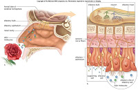
Figures 14.4 and 14.5 from the text show the taste and smell receptors.

 D. Sense of Vision
1. Anatomy and physiology of the eye
a. Table 14.2
b. function of the lens
i. cornea with lens and humors - focuses images on retina
ii. viewing near object - ciliary muscle contracts - tension released on suspensory ligaments - allows lens to round up
iii. viewing far object - ciliary muscle relaxed - suspensory ligaments taut - lens is flat
c. visual pathway to the brain
i. photoreceptors - absorption of light ->rod cells - rhodopsin splits into opsin & retinal - release of inhibitory molecules cease - signals go to other neruons in retina. ->cone cells - Blue, green, red pigments
ii. retina - rod & cone cells synapse with bipolar cells - synapse with ganglion cells whose axons become the optic nerve. many (150) rods active 1 ganglion. some cones activates 1 ganglion. integration occuring as signals pass to bipolar & ganglion
iii. blind spot
iv. from the retina to teh visual cortex - impulses from eyes along optic nerve to optic chiasma. fibers from rt 1/2 of each retina converge, continue in rt optic tract. fibers from left 1/2 of each retina converge, continue in left optic tract. fibers synapse with neurons in nuclei w/in the thalamus. nerve impulses to visual cortex
2. Abnormalities of the eye near & farsightedness, astigmatism
Figure 14.6 from the text shows the anatomy of the human eye. Additional images from the text can be found here.
 E. Sense of Hearing
1. Anatomy and physiology of the ear
a. outer ear - pinna, auditory canal
b. middle ear - tympanic membrane, oval & round window, ossicles (malleus, incus, stapes)
c. auditory tube
d. inner ear - semicircular canals & vestibules (equilibrium) & cochlea (hearing)
e. auditory pathway to the brain - tympanic memb. vibrates - to malleus, incus stapes (pressure multiplied x20). stapes strikes oval window - pressure to fluid in cochlea. movement of pressure waves from vetibular to tympanic canal across basilar membrane causes stereocilia of hair cells to bend. nerve impulses begin in cochlear nerve, travel to auditory cortext in temporal lobe for interpretation
Figures 14.13 and 14.14 illustrate the general anatomy of the ear and, more specifically, of the inner ear.

 F. Sense of Equilibrium
1. Rotational equilibrium pathway
a. mechanoreceptors in semicircular canals detect rotational equilibrium
b. displacement of cupula in ampulla causes stereocilia of hair cells to bend
c. pattern of impulses to brain changes
d. brain uses info to adjust motor output to right position in space
2. Gravitational equilibrium pathway
a. mechanoreceptors in utricle (back-forth movement) & saccule (up-down movement) detect movement of head in vert or horiz plane
b. movement causes displacement of otoliths, otolithic membrane sags, stereocilia of hair cells bend.
Click here for figure 14.15 from the text which shows the mechanoreceptors for equilibrium.
Definitions from Chapter 14 can be found here.
REFERENCES:
Mader, Syliva S. Human Biology. New York, NY: McGraw-Hill (2008).
Links provided throughout the summary take you to online sources.
IMPORTANT NOTE: Any time "text" or "the text" is referenced in the above summary, I am referring to the textbook Human Biology by Sylvia Mader (cited directly above).


No comments:
Post a Comment